A new paper, to which I collaborated while a postdoc in IPANEMA, is published Open Access today. Using an advanced synchrotron imaging method called Ptychographic X-ray Computed Tomography our international team of scientists from Brazil, France and Switzerland has obtained the most detailed 3D views ever achieved of very ancient traces of life on Earth. Lead by Dr. Lara Maldanis, this work investigates on the composition of 1.88 billion-year-old microfossils from Canada that are the preserved remains of microorganisms similar to bacteria existing today, but from a period when only microscopic life existed on Earth. The use (for the first time) of ptychography on such microfossils provided unprecedented details of their shape, composition and preservation. Surpisingly, in one locality, fossils previously described as “hematite-coated” are revealed to be composed of organic material – invisible in optical microscopy – coated with crystals of the iron oxide maghemite, instead of hematite. These findings challenge our understanding of past life and open exciting perspectives for the study of even older fossils or future samples returned from Mars.
For decades, scientists have been using fossils of microorganisms to better understand the origin and evolution of life on Earth, but this branch of palaeobiology has taken a great leap forward with the development of novel imaging techniques. Historically, the study of the earliest traces of life on Earth has been surrounded by a lot controversy and technical challenges. Sometimes it is even difficult to tell out if a structure is really a fossil or… just an artefact.
These challenges are related to the characteristics of these microfossils: they are only few micrometers in size (ten times less than the thickness of a human hair). Also, ancient rocks have suffered some degree of geological alteration due to the pressure and temperatures exerted by layers of rock above them. Therefore, the original components of these tiny cells have been “cooked” at temperatures of more than 100°C, substituted by minerals and pressed for hundreds of millions to billions of years, before ending up in the hand of scientists.
This challenge is shared with astrobiology in the search of life on other planets, like Mars. NASA and ESA are planning a mission for returning samples from that planet in the next decade to search for signatures of life, among other objectives. A main question is: if there are fossil microorganisms on Martian rocks, how are we going to identify them? To be ready for this task, we need to develop approaches to unequivocally identify the earliest traces of life on Earth and understand the conditions that allowed them to be preserved for so long.
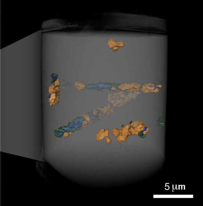
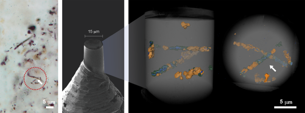
To study the fossil cells inside the rock without having to crack it open, our team used state-of-the-art high resolution Ptychographic X-ray Computed Tomography at the Swiss Light Source, Paul Scherer Institute. We were then able to reconstruct in 3D with 50-60 nm resolution the cells morphology, but also their chemistry as ptychography provides materials identification based on their electron density. Details obtained at this scale unprecedentedly inform on how time and geological processes have affected their original shape, which happens to be different in two localities of the same geological formation. In one locality, called Mink Mountain, the fossils had been previously described as hematite-coated structures, but we identified masses of mature carbonaceous materials that could not be seen using optical microscopy. Furthermore, instead of hematite, crystals of the less-common iron oxide maghemite are found associated with the fossils, revealing a previously unknown process involved in the preservation of these structures, and improving our comprehension of how these structures were preserved and how they were altered after being buried for billions of years.
This study also opens new and exciting possibilities for the controversies surrounding the oldest traces of life that have already been reported. Currently, synchrotron technology is taking a new step with the onset of 4th generation accelerators, such as SIRIUS, in Brazil, and MAX IV in Sweden, and the upgraded 3rd generation machines like the upgraded ESRF in Grenoble, and further SOLEIL, in Paris-Saclay, and the Swiss synchrotron SLS, both under preparation. These sophisticated machines have the potential to reveal further novel and exciting details about the earliest traces of life on Earth or even from Mars, and might help us answer some of the most intriguing questions of science: how did life appear on Earth? And are we alone on the Universe?
Reference: Maldanis L., Hickman-Lewis K., Verezhak M., Gueriau P., Guizar-Sicairos M., Jaqueto P., Trindade R.I.F., Rossi A.L., Berenguer F., Westall F., Bertrand L. & Galante D. 2020. Nanoscale 3D quantitative imaging of 1.88 Ga Gunflint microfossils reveals novel insights into taphonomic and biogenic characters. Scientific Reports 10: 8163. Find the article (Open Access) here