Born from an impropbable discussion at the Stanford synchrotron 3 years ago, my colleagues and I are publishing today in Science Advances (Open Access) a promising new technique for studying the chemical nature of carbon in 2D or 3D in entire organic fossils.
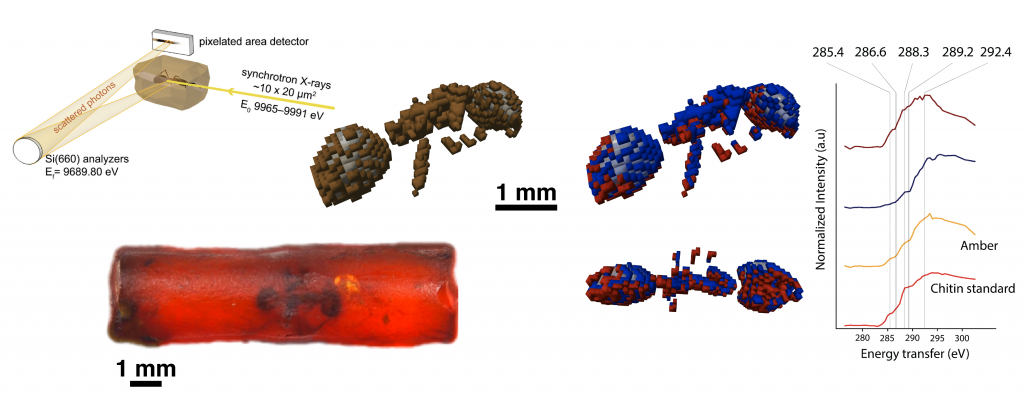
Our international team led by the IPANEMA laboratory (Paris-Saclay), and composed of three other CNRS laboratories (ISYEB, IMPMC and LRCP), the University of Lausanne and three synchrotron facilities (SOLEIL and ESRF in France, SLAC at Stanford in the United States) used synchrotron X-ray Raman scattering (XRS)–based imaging at the carbon K-edge to form 2D and 3D images of the carbon chemistry in a Carboniferous fossil trunk fragment and a 53-million-year-old ant trapped in amber.
The 2D XRS imaging of the trunk reveals a homogeneous chemical composition with micrometric ‘pockets’ of preservation, likely inherited from its geological history.
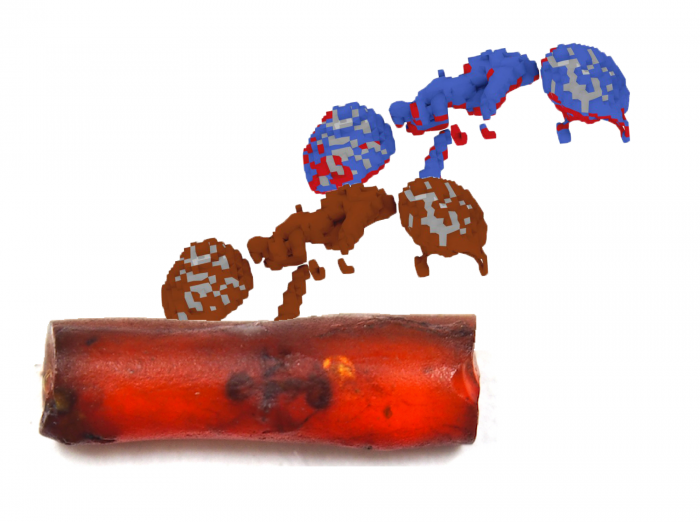
The figure above shows the 3D XRS imaging of the ant trapped in amber. Most of its exoskeleton (brown) displays an exceptionally well preserved remaining chemical signature typical of polysaccharides such as chitin, around a largely hollowed-out inclusion (grey). Although the achieved resolution is far from that of µCT, we could distinguish two degrees of preservation within the exoskeleton, the surface of the ant that was likely first brought into contact with resin (red) appearing better chemically preserved than the areas covered later after the ant’s death (blue). Watch a summary video here!
These results open up new perspectives for in situ chemical imaging of fossilized organic materials, with the potential to enhance our understanding of organic specimens and their paleobiology.
Reference: Georgiou R., Gueriau P., Sahle C., Bernard S., Mirone A., Garrouste R., Bergmann U., Rueff J.-P. & Bertrand L. 2019. Carbon speciation in organic fossils using 2D to 3D x-ray Raman multispectral imaging. Science Advances 5, eaaw5019. Find the article (Open Access) here

